Research Achievements
Bone’s drives scientific advancements in the field of orthopedic
medicine through in-depth research and technological innovation
Thoracic Vertebra Muscle Fat Infiltration and Degeneration Study
A correlation study on the degree of muscle fat infiltration and thoracic vertebra degeneration using Bone's self-developed QCT measurements. - Published in the international journal European Spine Journal (SCI Q1)
Proximal Femur Trabecular Bone Density Measurement Study
A comparative study of Bone's self-developed QCT and traditional QCT in measuring proximal femur trabecular bone density. - Published in the international journal Osteoporosis International (SCI Q1)
Bone Porosity Distribution Study
Innovative research results on bone porosity indicators for strength evaluation by Bone's Technology. - Published in the international journal Bone (SCI Q1)
| Year of Publication | Title | Journal |
|---|---|---|
| 2026 |
📄
Automatic phantom-less QCT predicts hip fracture risk: Bone and muscle parameters comparison
JCR Q1
This study evaluated the predictive ability of areal BMD (aBMD), volumetric BMD (vBMD), and intermuscular adipose tissue (IMAT) for hip fracture risk using an automatic phantom-less QCT (PL-QCT) system. Analysis of 550 patients showed that IMAT was significantly higher and aBMD/vBMD significantly lower in the fracture group (p < 0.001). Multivariate analysis identified IMAT as a risk factor and vBMD as a protective factor. IMAT outperformed aBMD and vBMD in predicting fracture risk (AUC: 0.896, 0.768, and 0.815), and their combination achieved the highest predictive value (AUC = 0.927). These findings highlight the importance of PL-QCT multiparameter measurements, especially IMAT, in improving hip fracture risk prediction and guiding clinical decisions.
Li, Wen, et al. European Journal of Radiology (2026)
|
European Journal of Radiology |
| 2025 |
📄
Validation of the Efficacy of Phantom-less Quantitative CT in Diagnosing Osteoporosis in Patients with Lumbar Degenerative Diseases[J]
北大核心 CSCD核心
This study evaluated the efficacy of phantom-less quantitative CT (PL-QCT) in diagnosing osteoporosis in patients with lumbar degenerative diseases. Data from 1,248 patients were analyzed, and the results showed that the osteoporosis detection rate of PL-QCT (50.2%) was significantly higher than that of spinal DXA (33.9%), hip DXA (39.1%), and dual-site DXA (43.2%) (P < 0.001). Using DXA as a reference, the sensitivity and specificity of PL-QCT were 79%-85% and 55%-81%, respectively, with an AUC of 0.75-0.82. The study indicates that PL-QCT has a higher detection rate of osteoporosis and demonstrates good diagnostic performance.
万文涛, 等. 中华骨科杂志(2025)
|
中华骨科杂志 |
| 2025 |
📄
Age- and sex-related changes in proximal humeral volumetric BMD assessed via chest CT with a deep learning-based segmentation model
JCR Q1
This study developed a deep learning-based method using nnU-Net to measure proximal humeral vBMD from chest CT scans and analyzed its age- and sex-related changes. The method showed excellent accuracy (DSC 98.42%, IoU 96.89%). In males, vBMD declined steadily from early adulthood, while in females, a sharp decline began around 40-45 years due to menopause-related bone loss. Lumbar spine vBMD decreased earlier than humeral vBMD, but their correlation was low to moderate, weakening after age 50. The findings highlight distinct sex-specific aging patterns and show that lumbar spine vBMD cannot replace proximal humeral vBMD in clinical assessments.
Li, Shihuai, et al. Osteoporosis International (2025)
|
Osteoporosis International |
| 2025 |
📄
Effects of lumbar degeneration on the global and regional bone mineral density in osteoporotic patients requiring lumbar interbody fusion for degenerative lumbar diseases JCR Q2
This study investigated the impact of lumbar disc degeneration (LDD) on bone mineral density (BMD). Analysis of 77 patients with lumbar degenerative diseases and osteoporosis showed that lumbar T-scores and endplate volumetric BMD (EP-vBMD) increased in lower lumbar segments and were significantly associated with LDD grading, while trabecular HU values were more sensitive to age-related systemic BMD changes and showed no strong correlation with LDD. The findings suggest that LDD significantly affects regional BMD, with EP-vBMD being closely related to local degeneration. Combining global and regional BMD assessments is recommended to improve diagnostic accuracy in patients with LDD.
Zhao, Yi, et al. European Spine Journal (2025)
|
European Spine Journal |
| 2025 |
📄
Quantitative computed tomography provides improved accuracy for diagnosis of lumbar osteoporosis in patients with facet joint osteoarthritis: a cross-sectional study JCR Q1
This study compared QCT and DXA in assessing BMD and their detection rates of lumbar osteoporosis in patients with facet joint osteoarthritis (FJOA). Analysis of 219 participants (mean age 65 years, 70.8% women) showed that QCT identified osteoporosis more frequently than DXA (58% vs. 34.2%, p < 0.0001). Severe FJOA was associated with lower vBMD on QCT but higher aBMD on DXA. As FJOA severity increased, aBMD measured by DXA rose, while vBMD measured by QCT declined. These findings indicate that QCT is more accurate for regional BMD assessment in FJOA patients, highlighting the need for caution when using DXA in clinical practice.
Wang, Xiayan, et al. Osteoporosis International (2025)
|
Osteoporosis International |
| 2025 |
📄
Adjacent Vertebral BMD Decline After Lateral Lumbar Interbody Fusion
JCR Q2
This study aimed to evaluate changes in the volumetric bone mineral density (vBMD) of cancellous bone and endplates adjacent to the fused vertebrae in LLIF patients and their relationship with mechanical complications. A retrospective analysis of 32 patients (mean age 60.1 years) showed that postoperative vBMD in all measured regions (UIV + 1, LIV + 1, UIV + 1e, LIV + 1e) was significantly lower than preoperative values (P < 0.05). While no significant differences were found between UIV + 1 and LIV + 1 in vBMD changes, endplate vBMD showed significant variation at follow-up (P = 0.035). These findings suggest QCT-based vBMD evaluation can provide insights into reducing mechanically related complications after LLIF surgery.
Sun, Kai, et al. Orthopaedic Surgery (2025)
|
Orthopaedic Surgery |
| 2025 |
📄
Using Phantomless QCT for evaluating BMD evolution in maintenance hemodialysis patients
JCR Q1
This study used phantom-less QCT (PL-QCT) to evaluate bone mineral density (BMD) changes in maintenance hemodialysis (MHD) patients. Thoracolumbar, total hip, and femoral neck BMD were measured in prospective and retrospective cohorts. Thoracolumbar BMD gradually decreased within the first 36 months of MHD but began to increase after 36–48 months, significantly exceeding pre-dialysis levels after 60 months. Hip and femoral neck BMD also decreased within 36 months but slightly increased after 72 months, returning to levels similar to the first year of MHD. It is recommended to use QCT to assess thoracolumbar or hip BMD during the first 3 years of MHD and focus on thoracolumbar BMD changes after 5 years.
Shen, Yuwen, et al. Scientific Reports (2025)
|
Scientific Reports |
| 2025 |
📄
Assessing vertebral bone density changes with phantomless QCT after posterior open reduction and internal fixation
SCI Q1
This study used a novel automatic phantom-less QCT (PL-QCT) system to evaluate short- and long-term vertebral BMD changes after posterior open reduction and internal fixation for thoracolumbar fractures. A total of 109 patients (70.6% male, median age 48) were included, with a median follow-up of 441 days. Patients were divided into Group 1 (378 days) and Group 2 (553 days). Postoperative BMD in all segments was significantly lower than preoperative values (P < 0.05), with no significant difference in BMD loss between the two groups (P > 0.05). BMD loss in the anterior region of the caudal adjacent vertebra was greater than in the posterior region (−13.0 mg/cm³ vs. −12.0 mg/cm³, P = 0.006). Early monitoring and intervention for postoperative bone loss, especially in the anterior region of adjacent vertebrae, is recommended to improve outcomes, requiring further follow-up studies.
Shao, Hu, et al. Scientific Reports (2025)
|
Scientific Reports |
| 2025 |
📄
Is Bone Mineral Density at the Tendon-Bone Interface After ACL Reconstruction Associated With Graft Maturation? A Quantitative Computed Tomography Analysis JCR Q1
This study investigated changes in bone mineral density (BMD) at the tendon-bone interface (TBI) after anterior cruciate ligament reconstruction (ACLR) and its association with graft maturity. Using phantom-less quantitative CT and MRI, BMD at femoral and tibial tunnel regions was measured in 36 patients over 2 years. BMD increased significantly within the first year (+36.1% to +57.5% at 6 months, +15.8% to +26.1% from 6 months to 1 year, P < .05) but showed minimal changes afterward (P > .05). Negative correlations were observed between BMD changes and graft maturity (SNQ values, r = -0.427 to -0.542, P < .05). These findings indicate that BMD at the TBI progressively increases within 2 years and can serve as a valuable reference for assessing tendon-bone healing
Lu, Wenhao, et al. The American Journal of Sports Medicine (2025)
|
The American Journal of Sports Medicine |
| 2025 |
📄
Automatic phantom-less calibration of routine CT scans for the evaluation of osteoporosis and hip fracture risk
JCR Q2
This study assessed the effectiveness of automatic phantom-less QCT (PL-QCT) in diagnosing osteoporosis and predicting hip fracture risk. Analysis of 705 patients showed that PL-QCT measurements strongly agreed with those of DXA and PB-QCT. PL-QCT demonstrated an AUC of 0.903 for diagnosing osteoporosis, comparable to PB-QCT, and an AUC of 0.869 for predicting hip fractures, outperforming DXA (AUC 0.831). Combining femoral neck BMD with inter-muscular adipose tissue percentage further improved prediction accuracy (AUC = 0.929). These findings suggest that PL-QCT is a reliable and valuable tool for clinical practice.
Li, Wen, et al. Bone (2025)
|
Bone |
| 2025 |
📄
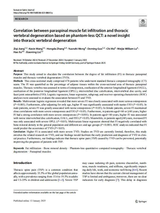
Correlation between paraspinal muscle fat infiltration and thoracic vertebral degeneration based on phantom-less QCT: a novel insight into thoracic vertebral degeneration JCR Q2
This study investigated the relationship between fat infiltration (FI) in thoracic paraspinal muscles and thoracic vertebral degeneration (TVD). Using advanced imaging techniques, FI was accurately measured and analyzed for its correlation with TVD severity. The results showed a positive association between FI and the severity of TVD, providing new insights for the prevention and treatment of thoracic degenerative diseases in clinical practice.
Jiang, Ziqi, et al. European Spine Journal (2025)
|
European Spine Journal |
| 2025 |
📄
Asymmetric distribution of vertebral bone microstructure in coronal adult spinal deformity: a cross-sectional study based on quantitative CT JCR Q2
This study used phantom-less quantitative CT (PL-QCT) to analyze asymmetric vertebral bone microstructure in adult spinal deformity (ASD) patients with coronal plane deformities, focusing on quantifying bone voids (BMD < 40 mg/cm³). Among 167 ASD patients, bone voids were predominantly located on the posterior-inferior concave side, with significantly higher volumes than the convex side. Female patients had greater bone void volumes than males. The findings indicate significant spatial heterogeneity and asymmetry in vertebral bone microstructure, influenced by abnormal biomechanical loading and remodeling imbalances. Quantifying bone void distribution provides insights into ASD progression and strategies to strengthen the concave side.
Ji, Chenqing, et al. European Spine Journal (2025)
|
European Spine Journal |
| 2025 |
📄
Biomechanical comparison of suspensory traction and axial traction in preoperative correction of cervical kyphosis: a finite element study
JCR Q1
This study compared the biomechanical characteristics of axial traction and suspensory traction in preoperative correction of cervical kyphosis using a C2-T2 finite element model. Both methods reduced the kyphotic angle, with suspensory traction achieving a greater correction (41° to 26° vs. 32° with axial traction). Suspensory traction shortened the C3-C7 spinal canal length (61.3 mm to 59.6 mm), while axial traction slightly increased it (to 61.8 mm). High stress areas were located at the anterior longitudinal ligament attachment in both methods, but suspensory traction resulted in lower overall stress. In conclusion, suspensory traction provides better correction of cervical kyphosis while reducing spinal canal length and vertebral stress, potentially lowering the risk of nerve damage and iatrogenic fracture.
Chen, Hongyu, et al. Frontiers in Bioengineering and Biotechnology (2025)
|
Frontiers in Bioengineering and Biotechnology |
| 2025 |
📄
Comparative analysis of endplate volumetric bone mineral density and endplate vertebral bone quality for predicting cage subsidence in lateral lumbar interbody fusion JCR Q1
This study compared QCT-based endplate volumetric bone mineral density (EP-vBMD) and MRI-based endplate vertebral bone quality (EBQ) scores in predicting cage subsidence (CS) after lateral lumbar interbody fusion (LLIF). Analysis of 97 patients showed that EP-vBMD was lower and EBQ higher in the CS group compared to the NCS group. EP-vBMD had better predictive efficacy than EBQ, with the highest AUC achieved by the combined model of EP-vBMD and EBQ (0.899), though not significantly better than EP-vBMD alone (p = 0.547). The findings suggest that regional endplate BMD assessments using QCT are more effective in predicting CS than MRI-based EBQ or global BMD measurements.
JCR Q1
Bian, Hanming, et al. Journal of Neurosurgery: Spine (2025)
|
Journal of Neurosurgery: Spine |
| 2024 |
📄
The novel phantom-less quantitative computed tomography for different CT machine systems: Bone mineral density quality assurance evaluation
JCR Q3
This study evaluated the quality assurance of a novel phantom-less QCT (PL-QCT) system for bone mineral density (BMD) measurements across 10 CT machines. Using the European spine phantom (ESP) as a reference, measurements showed high accuracy, with deviations within ±5mg/cm³ for most machines. One machine exceeded the threshold for L3 measurements. The calibration employed an automatic algorithm for soft tissue ROI placement, achieving a low coefficient of variation (~0.12%). The results highlight PL-QCT as a reliable and precise tool for osteoporosis evaluation, with potential for retrospective screening and wider clinical application.
Wei, Miao, et al. Journal of Medical Imaging and Radiation Sciences (2024)
|
Journal of Medical Imaging and Radiation Sciences |
| 2024 |
📄
Correlation Between Osteoporosis and Endplate Damage in Degenerative Disc Disease Patients: A Study Based on Phantom-Less Quantitative Computed Tomography and Total Endplate Scores JCR Q2
This study investigated the correlation between osteoporosis and vertebral endplate damage. Using advanced imaging techniques, the researchers assessed bone mineral density and endplate integrity in patients, revealing a significant association between osteoporosis and endplate damage. These findings are crucial for understanding the mechanisms of disc degeneration and developing strategies to prevent endplate damage.
Zhang, Yiming, et al. World Neurosurgery (2024)
|
World Neurosurgery |
| 2024 |
📄
Using automatic phantom-less quantitative computed tomography system based on routine CT to evaluate osteoporosis and predict hip fracture JCR Q3
This study validated the accuracy of a novel automatic phantom-less QCT (PL-QCT) for measuring bone density and assessed its value in diagnosing osteoporosis and predicting hip fractures. Data from 625 patients showed strong consistency between PL-QCT, DXA, and PB-QCT. PL-QCT achieved AUCs of 0.903 and 0.902 for diagnosing lumbar and hip osteoporosis, and 0.863 for predicting hip fractures. The study concludes that PL-QCT is better than DXA for predicting hip fractures and comparable to PB-QCT for diagnosing osteoporosis.
Li, Wen, et al. Journal of Medical Imaging and Radiation Sciences (2024)
|
Journal of Medical Imaging and Radiation Sciences (2024) |
| 2024 |
📄
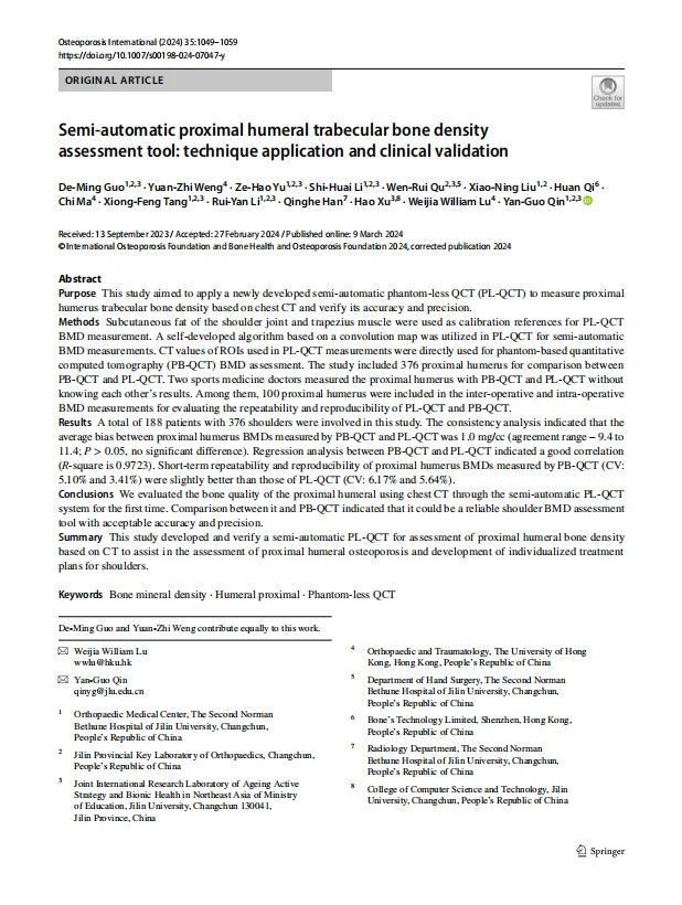
Semi-automatic proximal humeral trabecular bone density assessment tool: technique application and clinical validation JCR Q1
This study developed a semi-automatic phantom-less QCT (PL-QCT) tool to assess proximal humerus trabecular bone density based on chest CT. Compared to traditional QCT, the tool demonstrated high accuracy and efficiency. Results showed that it reliably measures proximal humerus bone density, providing a valuable tool for evaluating fracture risk and assisting in clinical assessments of proximal humeral osteoporosis.
Guo, De-Ming, et al. Osteoporosis International (2024)
|
Osteoporosis International |
| 2024 |
📄
Finite element analysis of a new preoperative traction for cervical kyphosis: suspensory traction
JCR Q2
This study established a finite element model of cervical kyphosis to analyze cervical spine stress under suspensory traction and its correction mechanism. A patient with C2-C5 cervical kyphosis underwent CT imaging for reconstructing C2-T2 vertebrae, followed by finite element analysis. Results showed that suspensory traction significantly reduced the kyphotic angle from 45° to 13°, primarily due to changes in kyphotic segments. Stress was concentrated in the anterior and posterior parts of the annulus fibrosus (except C4-5) and decreased in the anterior longitudinal ligament (ALL) from rostral to caudal. High stress was observed in C2-C5 segments, while other ligaments showed minimal roles. The correction was achieved through shear stress in the annulus fibrosus and tension in the ALL.
Chen, Hongyu, et al. Medical & Biological Engineering & Computing (2024)
|
Medical & Biological Engineering & Computing |
| 2023 |
📄
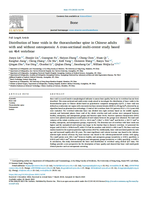
Distribution of bone voids in the thoracolumbar spine in Chinese adults with and without osteoporosis: A cross-sectional multi-center study based on 464 vertebrae JCR Q2
This study investigated the distribution of bone voids in the thoracolumbar spine using QCT. Results showed significant differences in bone void distribution across thoracolumbar regions, providing valuable insights into the mechanisms of vertebral fractures and guiding individualized prevention strategies.
Lin J, et al. Bone (2023)
|
Bone |
| 2023 |
📄
Performance evaluation of an AI-based preoperative planning software application for automatic selection of pedicle screws based on computed tomography images JCR Q2
This study evaluated an AI-based planning software for pedicle screw placement in spinal surgery. The software automatically recommended appropriate screw dimensions and positions based on imaging data. Results showed that AI-assisted planning significantly improved screw selection accuracy and surgical safety compared to freehand techniques.
Jia S, et al. Frontiers in Surgery (2023)
|
Frontiers in Surgery |
| 2023 |
📄
Cortical Endplate Bone Density Measured by Novel Phantomless Quantitative Computed Tomography May Predict Cage Subsidence more Conveniently and Accurately JCR Q2
This study used a novel phantom-less QCT (PL-QCT) technique to measure cortical endplate bone density and investigated its relationship with cage subsidence risk. The results showed that cortical endplate bone density is an important predictor of cage subsidence, providing a new assessment method for clinical prevention of cage subsidence.
Di M, et al. Orthopaedic Surgery (2023)
|
Orthopaedic Surgery |
| 2022 |
📄
One Novel Phantom-Less Quantitative Computed Tomography System for Auto-Diagnosis of Osteoporosis Utilizes Low-Dose Chest Computed Tomography Obtained for COVID-19 Screening JCR Q1
This study developed a novel phantom-less QCT (PL-QCT) system to measure bone mineral density (BMD) and diagnose osteoporosis using low-dose CT (LDCT) scans, such as those for lung cancer or COVID-19 screening. The system automates tissue selection and BMD calibration, improving precision and efficiency. Analysis of 649 LDCT scans showed the system accurately predicted osteoporosis (AUC: 0.68–0.88 compared to DXA) and tracked age-related BMD changes consistent with population trends. With strong correlations between CT values and BMD (r = 0.85–0.99), this PL-QCT system shows promise as a reliable tool for osteoporosis assessment in routine LDCT imaging.
Xiongfeng T, et al. Frontiers in Bioengineering and Biotechnology (2022)
|
Frontiers in Bioengineering and Biotechnology |
| 2022 |
📄
Patient-specific numerical investigation of the correction of cervical kyphotic deformity based on a retrospective clinical case
JCR Q1
This study used patient-specific numerical simulation methods to investigate cervical kyphosis. By constructing individualized finite element models, the researchers simulated the effects of different degrees of cervical kyphosis on spinal cord stress, providing a theoretical basis for clinical treatment decisions.
Wu T, et al. Frontiers in Bioengineering and Biotechnology(2022)
|
Frontiers in Bioengineering and Biotechnology |
| 2022 |
📄
Endplate volumetric bone mineral density biomechanically matched interbody cage
JCR Q1
This study highlights that endplate volumetric bone mineral density (EP-vBMD), measured using automatic phantom-less quantitative CT, is an effective predictor of cage subsidence and a guide for cage selection in interbody fusion surgery. Porous metallic cages have become a promising solution to reduce subsidence risk while retaining the superior osseointegration capabilities of titanium alloy cages. Customizing the elastic modulus of porous metallic cages based on patient-specific EP-vBMD allows for biomechanical matching with the contacting bone tissue. A novel customization method is proposed, adjusting cage porosity to modify its elastic modulus, with a three-grade porosity strategy or direct customization available to meet patient or physician preferences.
Weng, Yuanzhi, et al. Frontiers in Bioengineering and Biotechnology (2022)
|
Frontiers in Bioengineering and Biotechnology |
| 2022 |
📄
A novel surgical planning system using an AI model to optimize planning of pedicle screw trajectories with highest bone mineral density and strongest pull-out force JCR Q1
This study evaluated a novel artificial intelligence (AI) model for optimizing transpedicular screw trajectories in osteoporotic patients to achieve higher bone mineral density (BMD) and pull-out force (POF). Using the Bone's Trajectory system, preoperative CT scans of 21 patients were analyzed. The AI model automatically identified alternative trajectories, calculated trajectory BMD, and estimated POF for L3-5. Compared to AO standard trajectories, the optimized trajectories had significantly higher BMD and POF (p < 0.05), with POF averaging at least two times higher. The findings demonstrate that the AI model effectively selects superior screw trajectories, outperforming traditional AO paths.
Ma, Chi, et al. Neurosurgical Focus (2022)
|
Neurosurgical Focus |
| 2022 |
📄
Automatic phantom-less QCT system with high precision of BMD measurement for osteoporosis screening: Technique optimisation and clinical validation
JCR Q1
This study validated the accuracy and precision of a newly developed automatic phantom-less QCT (PL-QCT) system for measuring spinal bone mineral density (BMD) and diagnosing osteoporosis. In 36 patients, BMD results from DXA and QCT were compared, while 63 patients underwent analysis with both PB-QCT and the new PL-QCT system. The automatic PL-QCT system showed higher precision than traditional QCT and comparable diagnostic performance to DXA and PB-QCT. ROC analysis (AUC > 0.8), strong correlation (r = 0.99), and Bland-Altman analysis demonstrated high accuracy and consistency. Automatic region of interest (ROI) selection improved precision (CV = 0.89%) and convenience. This system offers significant potential for osteoporosis diagnosis, translational research, and clinical application.。
Liu, Zhuo-Jie, et al. Journal of orthopaedic translation (2022)
|
Journal of Orthopaedic Translation |
| 2022 |
📄
Focal osteoporosis defect is associated with vertebral compression fracture prevalence in a bone mineral density-independent manner
JCR Q1
This study introduced focal bone mineral content (BMC) loss as a measure of focal osteoporosis defects and evaluated its link to vertebral compression fracture (VCF) prevalence. Analysis of 205 cases showed that focal BMC loss was highly reproducible, correlated well with bone microarchitecture (e.g., bone volume fraction), but weakly with trabecular BMD. Focal BMC loss was higher in the fracture group and independently associated with VCF risk. These findings highlight focal BMC loss as a reliable indicator of osteoporosis-related fracture risk, measurable using routine CT scans.
Li, Chentian, et al. JOR spine (2022)
|
JOR spine |
| 2020 |
📄
Automatic Measurement of Subregional Vertebral Bone Mineral Density via Deep Learning of Quantitative Computed Tomography Images JCR Q3
This study developed and validated a deep learning-based segmentation method for automatic subregional Bone Mineral Density (BMD) measurement using Quantitative Computed Tomography (QCT) scans. The method demonstrated high accuracy and reproducibility in vertebral body subregion segmentation, including cortical and cancellous bone, achieving precise BMD measurements. The automated framework provides a reliable and efficient solution for investigating subregional osteoporosis changes, enhancing clinical practice with QCT spine scans.
Li, Chentian, et al. International Journal of Orthopedics and Rehabilitation (2020)
|
International Journal of Orthopedics and Rehabilitation |